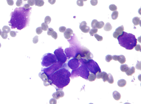
offers a safe, simple way of obtaining highly reliable samples. Since material is excised instead of being aspirated, complete cell clusters are obtained rather than single cells. As a result, at least 95% diagnostic accuracy has been obtained using Rotex for sampling lung tumours (1) and non-palpable breast tumours (2).
In a comparative study, the diagnostic yield for thyroid nodules was significantly better for the Rotex Screw Needle than for conventional fine needle cytology (97% vs. 83%, respectively; p<0.001) (3).
The diameter of the instrument cannula is 0.8, which explains the low incidence of reported complications (1).
|
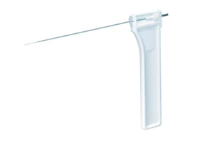
Construction of the instrumentThe screw needle is made of a 0.55 mm thick stainless steel rod with a small handle at its proximal end. The distal 16 mm of the rod have been formed into a tapering screw with cutting ridge. The needle is housed in a steel cannula with an outer diameter of 0.8 mm (equivalent to 21 needles). Read more »
|
Fields of applicationThe Rotex instrument was originally constructed for the biopsy of lung lesions. It has, however, proven to be equally suitable for the biopsy of other organs such as the liver, the kidney, lymph nodes, mammary tissue, thyroid etc. Even malignant bone lesions may be biopsied with the instrument. Read more »
|